mistory
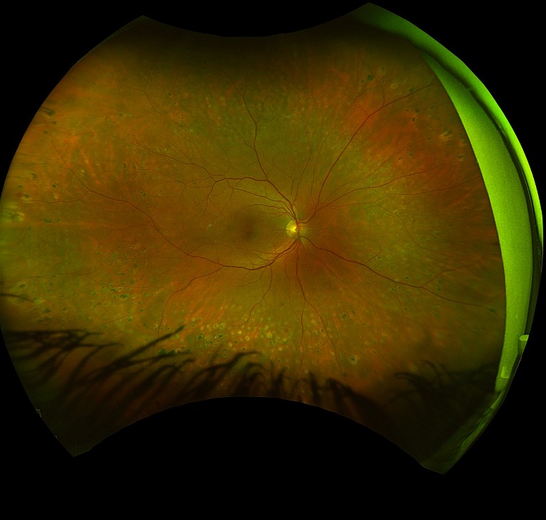
Proliferative diabetic retinopathy post PRP.
Revolutionising Diabetes Management

Ultra-widefield Retinal Imaging
As an eye care professional with a special interest in diabetes-related eye disease for almost 20 years and, having lived with type 1 diabetes for over 32 years, Dr Amira Howari’s journey has been deeply intertwined with a commitment to bridging the gap in our healthcare system. Throughout her career, she has become increasingly determined to help build a robust, holistic, and collaborative multidisciplinary approach to diabetic ocular care.
WRITER Dr Amira Howari
Navigating the intricate landscape of diabetic ocular care demands a holistic approach that integrates clinical acumen with cutting-edge technology.
With the global prevalence of diabetes reaching unprecedented levels, particularly in countries like Australia where diabetic retinopathy ranks as the leading cause of blindness among working-age adults,1,2 the demand for advanced diagnostic and management strategies has never been more pressing.
In my daily practice, I encounter patients at various stages of diabetic retinopathy, each presenting unique challenges in terms of diagnosis, monitoring, and treatment.
Traditional retinal imaging methods have served as the cornerstone of our diagnostic approach, providing valuable insights into retinal health.3 However, these methods are limited by their inability to capture a comprehensive view of the retina, especially in cases where pathology extends beyond the central macula. Despite this, I previously held the view that retinal scans were somewhat comparable, regardless of the technology used; after all, a scan is a scan.
And then, along came ultra-widefield (UWF) retinal imaging.
A WATERSHED MOMENT
The unveiling and launch of UWF retinal imaging technology in Australia marked a watershed moment in my professional journey. I was fortunate enough not only to operate a newly launched cuttingedge technology but also to undergo a demonstration as a patient. What truly captivated me, however, was not merely its operational simplicity, but the revelation it offered: the chance to witness my own retina with an unprecedented level of clarity, detail, and scope.
Despite years of comprehensive explanations from esteemed retinal ophthalmologists and colleagues, nothing prepared me for the sheer impact of seeing my retinal landscape unfold before my eyes. Seeing my retinal anomalies with such magnification and resolution was profound. The concept that had been explained to me suddenly materialised into a tangible reality on the screen. Though my visual acuity remained unaffected, the subtle changes observed in my retina over time left indelible impressions. They were timely reminders of the silent yet potent effects of diabetic retinopathy.

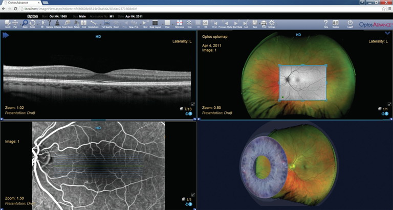
OptosAdvance automatically arranges modalities according to the individual user preference.
This transformative experience catalysed a significant shift in my perspective, both personally and professionally. It ignited within me a renewed dedication to the meticulous management of my own diabetes, as well as a heightened awareness of the importance of regular specialist appointments. Moreover, it fundamentally altered my approach to every subsequent diabetes eye consultation, from the routine task of history taking to the nuanced discussions surrounding diabetes management, and the painstaking examination of the retina.
As a clinician living with type 1 diabetes, it also triggered an innate responsibility to challenge the status quo and redefine the conventional norms taught in academic settings – a commitment that continues to drive my professional ethos to this day.
THE FUNDAMENTAL ADVANTAGES OF UWF
Ultra-widefield retinal imaging technology has revolutionised the way we approach diabetic eye care, offering a panoramic view of the retina in a single, non-invasive scan of up to 200° or more, encompassing up to 82% of its surface area.6,7
This expanded view enables better visualisation of peripheral retinal pathology, which is often overlooked with traditional imaging methods. As a clinician, this enables us to better identify and monitor subtle retinal changes in areas previously inaccessible with conventional imaging techniques.
Additionally, UWF retinal imaging devices significantly enhance the patient experience during retinal examination, due to the nonmydriatic imaging feature.8
Although this does not remove the need for dilation upon indication, it certainly serves to capture a comprehensive view of the retina without the need of dilation in many routine cases, eliminating the associated discomfort and inconvenience. This feature also increases compliance with regular screening protocols, ultimately leading to better long-term outcomes.
The patient experience is also enhanced with UWF technology’s speed and ease of use. The ability to capture high-quality images quickly, often within seconds, streamlines the imaging process and minimises patient wait times. This efficiency is further enhanced by the intuitive interface system, which allows for integration into the daily workflow.
From a clinician’s perspective, one of the most significant advantages of UWF retinal imaging is its impact on patient education and engagement. Being able to visually demonstrate retinal pathology on widefield images has proven invaluable in helping patients understand the significance of their condition and the importance of regular eye examinations. The ability to share these images with patients fosters open communication and empowers them to take an active role in managing their eye health.
Undeniably, the documentation and tracking capabilities of UWF retinal imaging have also revolutionised the way we monitor disease progression and treatment outcomes. The digital storage and easy accessibility of retinal images enable clinicians to track changes in retinal health over time, facilitating more informed decision making and personalised treatment plans for each patient.
“ Ultra-widefield retinal imaging technology has revolutionised the way we approach diabetic eye care ”
In recent years, UWF retinal imaging has been the focus of extensive research and development aimed at optimising its capabilities and clinical utility. Studies have highlighted its effectiveness in detecting diabetic retinopathy and other retinal pathologies with high sensitivity and specificity.9,10 Additionally, advancements in imaging technology have led to improvements in image resolution, allowing for even greater detail and accuracy in the visualisation of retinal structures.
The accompanying software of UWF retinal imaging devices has also been instrumental in enhancing the ability to evaluate and analyse retinal images effectively. With features such as zoom functionality, measurement tools, image enhancement filters, annotation tools, comparative analysis, and export and sharing options, the software streamlines the interpretation process and ensures accurate and comprehensive assessments of retinal health.
THE POWER OF CONSISTENCY
When optometrists and ophthalmologists comanage patients with diabetic eye disease, it is important that they use the same technology because consistency in retinal imaging has never been more critical for early detection and comparing results over time.
When different devices are used, we find that optometrists and ophthalmologists will repeat the assessment to ensure reliability, repeatability, and consistency when comparing results over time. This considerably adds to the burden of care for patients, their carers, the providers, and economy.
Ultra-widefield retinal imaging has transformed the delivery of diabetic ocular care. With ongoing advancements in research and technology, UWF retinal imaging will continue to support eye care professionals in their efforts to prevent vision loss for these patients.
Dr Amira Howari BOptom (Hons) GradCertOcTher MOptom (UNSW) is a senior clinical optometrist; healthcare industry and motivational keynote speaker; a Diabetes Australia and KeepSight Ambassador; and former Optometry Australia Councillor (NSW/ACT).
Dr Howari has worked in corporate, independent, ophthalmology, and pharmaceutical settings; and at the University of New South Wales as a guest lecturer and clinical supervisor. She currently serves as a member of the Diabetes and Endocrine Network – Agency of Clinical Innovation (NSW Health).
References
1. 2019 Global Burden of Disease Collaborative Network. Global Burden of Disease Study 2019. Seattle, United States: Institute for Health Metrics and Evalu.ation (IHME), 2020.
2. Tan, C.S., Chew, M.C., Lim, L.W., Sadda, S.R., Advances in retinal imaging for diabetic retinopathy and diabetic macular edema. Indian J Ophthalmol. 2016 Jan;64(1):76–83. DOI: 10.4103/0301-4738.178145.
3. Shoughy, S.S., Arevalo, J.F., Kozak, I. (2015). Update on wideand ultra-widefield retinal imaging. Indian Journal of Ophthalmology. 63. 575–81. DOI. 10.4103/0301-4738.167122.
4. Optomap ultrawidefield advances. Available at optos. com/globalassets/public/optos/providers/clinical-papers/css-uwfadvances-us.pdf.
5. Nanegrungsunk, O., Patikulsila, D., Sadda, S.R., Ophthalmic imaging in diabetic retinopathy: A review. Clin Exp Ophthalmol. 2022 Dec;50(9):1082–1096. DOI: 10.1111/ ceo.14170.
6. Ellery, B., Milverton, J., Merlin, T., Retinal photography with a non-mydriatic retinal camera in people with diabetes. MSAC application no. 1181 Assessment Report; 2014.
Commonwealth of Australia, Canberra, ACT.
7. Optos. (2023). Optos Ultra-Wide Field Imaging Software Manual.
8. Piyasena, M.M.P., Murthy, G.V.S., Kamalakannan, S., et al., Systematic review and meta-analysis of diagnostic accuracy of detection of any level of diabetic retinopathy using digital retinal imaging. Syst Rev. 2018 Nov 07;7(1):182. DOI: 10.1186/s13643-018-0846-y.
9. Hassan, B., Raja, H., Hassan, T. et al. A comprehensive review of artificial intelligence models for screening major retinal diseases. Artif Intell Rev 57, 111 (2024). DOI: 10.1007/s10462024-10736-z.