mieducation
Visual Snow Syndrome: Revealing the Mysteries
Visual snow (VS) is a recently accepted and increasingly researched condition where children and adults see pixelated dots in all of their vision; like an old TV screen with static. It may be present from a very early age, or it can be acquired at any age, sometimes in association with concussion or a health issue, but often there is no obvious reason for the onset of constant visual dots. Until recently, the symptoms were confused with migraine aura, but the conditions are separate.
VS may occur without other symptoms or with a range of symptoms, and the condition can be mild or so severe that the person’s ability to function is significantly affected. The pathophysiological cause is unknown, and there is no cure for VS, but evidence is evolving to suggest tinted lenses and/or optometric vision therapy may significantly reduce the symptoms in some people with visual snow.
WRITERS Stephen Leslie and Elizabeth Wason
LEARNING OBJECTIVES
On completion of this CPD activity, participants should be able to:
1. Be aware of visual snow and its prevalence,
2. Understand the symptoms and how it affects vision and quality of life,
3. Be aware of how the impact of visual snow may be reduced, and
4. Realise the role of optometric vision therapy in treating these patients.
Visual snow is a neurological condition in which people see pixelated moving black and white or coloured dots, usually constant, and over all of their vision.
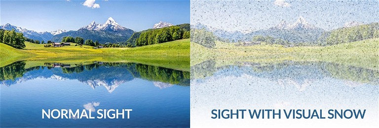
Sometimes described as visual noise (Figure 1), VS can be present from a very early age, and often children do not report it to parents or health care professionals as they have always seen it that way. Some children and adults may have minimal symptoms and effects on their activities of daily living and quality of life (QoL). Others can have major symptoms that severely limit their ability to function, use screens, drive at night, socialise (especially at night), work, and enjoy life.1,2
There may be no obvious cause for visual snow, but it is not uncommon following concussion,3,4 which is defined as a mild acquired brain injury. Usually benign, VS may be associated with a range of conditions, including some medications; trauma; stress; seizures; psychiatric disorders; ocular disease such as optic neuropathy or papilloedema; idiopathic intracranial hypertension (IIH); transient ischaemic attack (TIA); a history of cerebrovascular disease; or autoimmune disorders including Crohn’s disease, rheumatoid arthritis, and multiple sclerosis. Due to the common co-occurrence of a range of other symptoms described further on in this article, many cases of VS are referred to as visual snow syndrome (VSS), a term first used in 2013.5
However, the condition now called visual snow was first described in 1944 when the side effects of the drug digitalis for treatment of heart conditions were published.6 The appearance of multiple black and white coloured dots in the complete visual field has been confused in the past with migrainous aura, until Liu et al. described 10 patients diagnosed with migraine who had constant visual snow rather than episodic migrainous aura.7
Varying levels of prevalence of visual snow syndrome have been suggested, with Kondziella et al., in a United Kingdom study, reporting “2%, with another 2% having visual snow without meeting the criteria for visual snow syndrome”.8,9
DIAGNOSTIC CRITERIA
The Visual Snow Initiative (VSI, visualsnowinitiative.org) was founded by Sierra Domb, who has VS, and her family to increase awareness of the condition, to encourage and fund ongoing research, and to provide resources and support for people with VSS.

Figure 1. Comparison of normal and visual snow vision.

Figure 2. Palinopsia zebras.

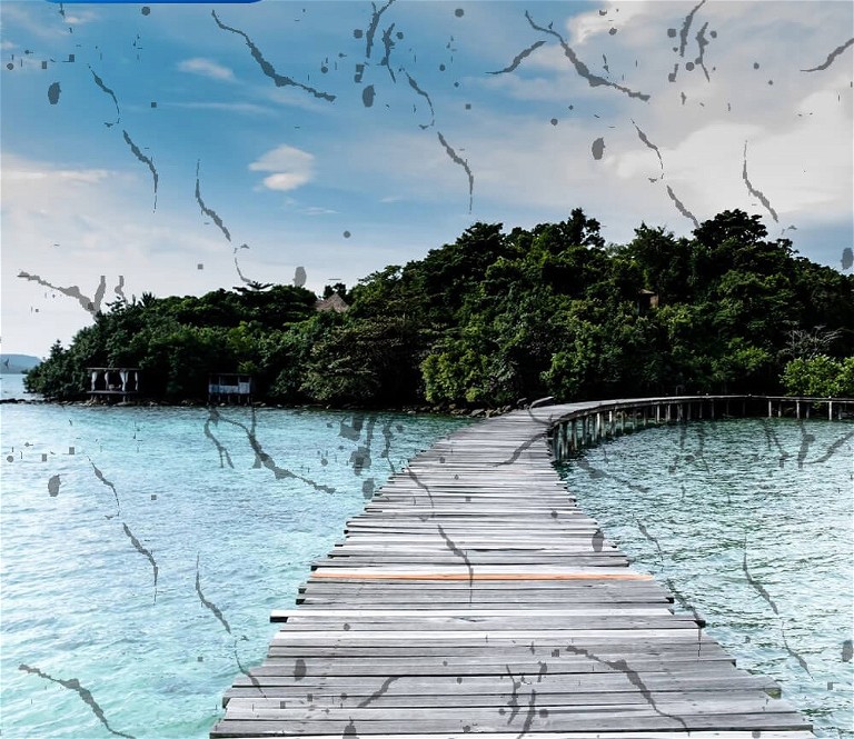
Figure 3. Multiple floaters.
The VSI has publicised diagnostic criteria for VS and VSS, namely: The dots should persist for at least three months, and be accompanied by at least two of the following:
1. A persistent after-image of an object or a comet-like tail behind a moving object, known as palinopsia (Figure 2),
2. Excessive floaters or coloured waves with eyes open or closed (Figure 3),
3. Significant sensitivity to light, 4. Impaired night vision, and
5. Symptoms that are not typical of a migraine aura or other diagnosis.
As well as visual symptoms, people with VSS may also experience non-visual symptoms such as:
• Ringing in the ears (tinnitus),10
• Depersonalisation (feeling detached from oneself at times),
• In some cases, anxiety and/or depression, and
• Tremor.
People with VS may also have migraines or brain fog; dizziness and/or nausea; sleep disorders; and tingling and/or pain in body parts.
It is important to ensure that the symptoms of visual snow are consistent with the diagnostic criteria for VSS, and there is an absence of ocular disease or other neurological symptoms or conditions, before a diagnosis of VSS is established.11-15 While many people with VS or VSS can also experience migraine, the features of VSS make it a different clinical entity to migraine.16-19
Children may not report visual snow to their parents or health care practitioners; one recent seven-year-old patient, who had previously visited five other health care practitioners, described her vision as “raindrops everywhere”.
As awareness of visual snow is still growing, it is not unusual for people with VS symptoms to experience health care practitioners who are unaware of the symptoms, and/or deny it exists, with patients often reporting that their symptoms have been dismissed or suggested as fictional. The condition may be misdiagnosed as migraine or other conditions, or not considered in differential diagnosis of a range of unusual symptoms. It is not uncommon in practice for patients provided with a diagnosis of VSS to report great relief following an explanation of their symptoms, and in some cases they are content to live with the mild effects and do not require any treatment.
VSI is taking actions to establish an International Classification of Diseases (ICD) code for VS and VSS, primarily to achieve a recognised medical status for the conditions.
PALINOPSIA
Illusory palinopsia, also known as visual perseveration, appears as a tail to a moving object, or a series of images of the object as it moves through space. The images are poorly formed, short-lasting, and unclear, as compared to the longer-lasting hallucinatory palinopsia, which is usually a clear and distinctly formed image, indicative of hallucinogenic persisting perception disorder (HPPD) due to use of LSD or magic mushrooms. While it is common with VSS, illusory palinopsia can be associated with many other conditions.
Patients with VSS and palinopsia can experience severe difficulties reading as the saccadic eye movements result in images of the previously read word overlying the nextfixated word, plus the added disturbance of visual snow dots. On computers, the visual snow dots interfere with reading the text on a lit background. The combination of visual snow pixels, photophobia, and palinopsia with trailing images of moving objects can make driving very difficult, especially at night with streetlights in the dark.20
PATHOPHYSIOLOGY OF VISUAL SNOW
The neurological explanation for visual snow is not completely known. A number of complex investigations have been reported, including ultra-high-field morphological and quantitative magnetic resonance imaging (MRI),21 which found significant correlations between MRI variables and VS symptoms and intensity of visual snow, but further research is needed to confirm these results. Research has suggested the condition may affect a number of brain areas that are involved with visual processing, including the temporal and parietal lobes.22-24 Aeschlimman et al. recently summarised the literature to date as “electrophysiological studies have revealed cortical hyperresponsivity in visual brain areas, imaging studies (have) demonstrated microstructural and functional connectivity alterations in multiple cortical and thalamic regions and investigated glutamatergic and serotoninergic neurotransmission”.25 Neuro-ophthalmologist Dr Gordon Plant recently published a summary of the current understanding of the visual snow pathophysiology online.47
VISUAL DYSFUNCTIONS
Accommodation, binocular vision, convergence, and eye movement dysfunctions have been found to be present in up to 60% of people with VSS,26-28 significantly more than in a general optometric population,29 with confirmatory laboratory studies of saccadic eye movements co-authored by Dr Allison McKendrick, now Chair of Optometry Research at University of Western Australia and the Lions Eye Institute.30-32
TREATMENT
Pharmacological treatments, including lamotrigine, valproate, topiramate, acetazolamide, and flunarizine, have been found to be usually unsuccessful, with adverse side effects in half of the patients treated, and may in fact increase the primary visual snow symptom.33
As yet, there is no complete cure for visual snow. However, two visual treatments have been found to significantly reduce the visual symptoms of VS in a number of case studies, providing improved ability to function in activities of daily living such as work, and improved QoL.
Chromatic Filters (Tinted Lenses)
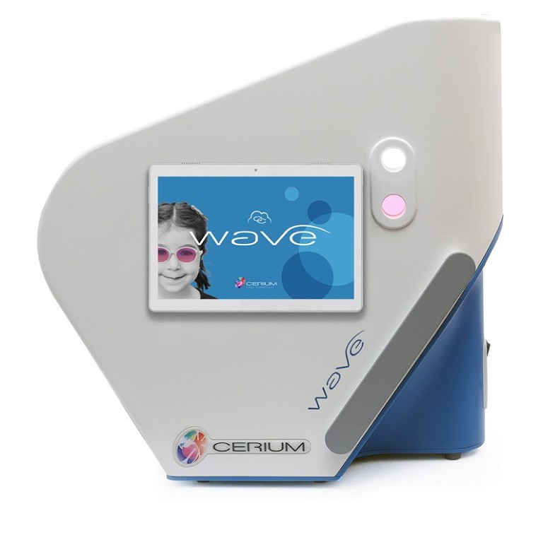
Sydney neuro-ophthalmologist Dr Clare Fraser and her colleagues Dr Jenny Lauschke and Dr Plant assessed a small number of subjects with visual snow symptoms using the Cerium Intuitive Colorimeter instrument (Figure 4). For this assessment, patients looked at a page of print in a closed instrument while the colour of the light was changed. The patient was asked to report whether each hue made the print clearer or blurred, moving or double, and if the light made them feel uncomfortable or nauseous, or more comfortable. If a particular illumination hue and density was determined to be most beneficial, software was used to calculate a combination of tinted lenses, which could be trialled for the patient under fluorescent lights, in normal lighting inside and outside, and looking at screens and reading, to assess the improvements in visual function and visual comfort.35,36 The instrument was developed by Professor Arnold Wilkins of the University of Essex to assess abnormal photophobia, which he calls visual stress, but is more commonly known as pattern glare.37-45 Dr Fraser prescribed tinted lenses, which reduced symptoms in most of the 32 patients studied.46-47 They suggested that visual snow was due to a “thalamocortical dysrhythmia of the visual pathway” and that the coloured filters “may alter the koniocellular pathway processing, which has a regulatory effect on background electroencephalographic rhythms”.

Figure 4. Intuitive Colorimeter instrument by Cerium.
A number of other studies have reported improvements in symptoms and visual functions with chromatic filters.48,49 Optometrists Dr Ken Ciuffreda and colleagues at the State University of New York (SUNY ) assessed more than 100 patients with VS using structured test protocols and found that 90% of the subjects (similar to Fraser et al.) preferred a specific-coloured filter, which was prescribed for constant wear and/or near visual tasks such as computer use and smartphones.48 Tannen et al. provided a retrospective analysis of optometric management of 27 patients diagnosed with VSS for whom they combined an individual prescription of chromatic tints (BPI and FL-41), with optometric vision therapy specifically for the accompanying palinopsia and eye movement issues. They reported symptom reduction of at least 50% with prescription of chromatic filters, and reduction of the palinopsia by 50–65% in the 23 patients who underwent vision therapy.
In practice, optometrists who employ the Cerium Intuitive Colorimeter to assess the potential benefits of tinted lenses, and to determine the specific hue and density, find that for some patients there is no preference for a particular light hue on the Intuitive Colorimeter, and tinted lenses make no difference to the visual symptoms, particularly for those who have had the condition for years. And yet for others in whom the condition is less chronic, specific chromatic filters often produce a dramatic reduction in visual symptoms, as well as major improvements in their ability to function in many activities of daily living.
The preferred chromatic filter can be very individual, with some preferring FL-41 (originally designed to treat sensitivity to fluorescent lights), a reddish lens that blocks 80% of blue light transmission in the spectrum of 480–520 nm. Unfortunately, FL-41 tinted lenses have become so commercialised online for migraine and other conditions that the actual tint and filtering characteristics can vary dramatically. At the same time, some patients with VSS find FL-41 tinted lenses very uncomfortable, and prefer a different hue, as determined by colorimetry assessment.
Neuro-Optometric Rehabilitation Vision Therapy
Neuro-optometric rehabilitation vision therapy has been found to improve QoL questionnaire scores, as well as distance and near visual activities, and social functioning.
Optometrists Dr Charles Shidlofsky of Texas, Dr Terry Tsang of California, and Visual Snow Initiative author Vanessa Mora collaborated in the first study of optometric assessment and vision therapy treatment of visual snow and associated visual and ocular conditions.50,51 They used the National Eye Institute Vision Function Questionnaire (NEI-VFQ-25) to measure QoL prior to and following neuro-optometric rehabilitation therapy in a pilot study of 21 people with VSS. The questionnaire was repeated after six and 12 weeks of vision therapy. All participants had comprehensive standard optometric evaluations of vision function.
Optometric vision therapy was individually programmed to improve eye movements, accommodation, and vergence functions where abnormal, and visual processing abilities.52,53 Each patient underwent 12 one hour in-office sessions of three to five vision therapy activities to develop improved visual skills, with home therapy to develop automaticity of the higher-level visual abilities.
Study participants showed statistically significant improvements in QoL scores at intervals of six and 12 weeks of vision therapy, and questionnaire subscales of distance and near vision function, “social functioning, mental health, role difficulties, and dependency also showed improvement”. However, the authors acknowledged the study’s potential limitations, recommending larger and more controlled studies would be necessary to assess the possible benefits of neuro-optometric vision therapy in improving visual functions, reducing symptoms, and improving QoL in people with VSS. Since this study was conducted, a scale to measure visual snow symptom severity has been developed.54
CASE ONE
Emily* is a 23-year-old medical typist referred by an optometrist to one of the authors. She reported visual snow dots in all of her vision for as long as she could remember. Her symptoms had increased significantly in the past two years, coincident with the onset of headaches every day. She denied aura and neurological company. Emily was being treated by a neurologist with regular botox scalp injections for her constant and severe headaches. Previous treatment with fluoexetine had been terminated due to side-effects, and she had experienced similar issues with the migraine medication galcanezumab. She experienced persistent after-images of lights and objects, and tails to moving objects, such as a waving hand or thrown ball, typical of illusory palinopsia. She also reported many floaters and coloured waves in her vision, even with her eyes closed; as well as significant photophobia and difficulties at night, especially with streetlights interfering with her ability to drive. When further questioned, she reported tinnitus, occasional depersonalisation, anxiety, infrequent dizziness, and variable sleep issues. She also had significant visual discomfort in shopping centres and found scrolling print on a screen very uncomfortable; she had turned the brightness of screens to the lowest level possible. She denied double vision but reported objects and people’s faces often seemed “ghosted”. Prism glasses prescribed elsewhere made the symptoms worse. Her mother had a history of migraines.
Emily had unaided distance visual acuities of 6/6 right and left, and no significant refractive error. Optometric assessment revealed depressed stereopsis at 400 arc seconds (random dot stereopsis), accommodative insufficiency, infacility and fatigue; and pain on convergence testing at 20 cm, which improved to 5 cm with trial of +0.50 sphere (both eyes), indicating a convergence dysfunction secondary to an accommodative dysfunction. Her pursuit and saccadic eye movements were mildly below normal levels for her age. She experienced moderate visual discomfort looking at the Wilkins Pattern Glare #2 test plate for visual stress/pattern glare. Her ocular health examination was unremarkable.
Intuitive Colorimetry testing and an indicated lens trial of a purple and rose tint, with 32% light absorption, provided significantly increased visual comfort. When trialled on a computer screen, she reported dramatically better clarity and comfort looking at the screen under fluorescent lights. Ghosting of objects was almost eliminated, as was object ghosting and trails of her moving hand.
Emily was informed in detail about visual snow syndrome and palinopsia, and prescribed a low plus prescription with a chromatic filter, to be worn for all near visual tasks. She was scheduled for review in six weeks for assessment of the improvement in symptoms, and to commence optometric vision therapy to normalise her visual functions. A report was provided for her neurologist and GP.
CASE TWO
Denny* is a 51-year-old phlebotomist who started seeing black and white dots in all of her vision, six months before she presented for optometric care. She had blurred and ghosted distance vision, but she denied any history of head injury or whiplash. She experienced frequent headaches but denied aura. She was on a disability pension for post-traumatic stress disorder, but did not have sleep issues. MRI investigation was unremarkable, and she was being treated for depression with desvenlafaxine (Pristiq, among others). Her sister was considered a glaucoma suspect, but there was no family history of migraine. Her ocular health was notable for moderate evaporative dry eye, bilateral posterior vitreous detachment and vitreous traction. She reported moderate brain fog and tinnitus but denied any episodes of depersonalisation. She reported mild tails to moving objects and showed a strong adverse reaction to the Wilkins visual stress/pattern glare test.
Questioning confirmed she met the diagnostic criteria for VSS, with black and white dots over all of her vision present for more than three months; mild palinopsia; increased entoptic phenomena; increased photophobia; severe difficulty at night with lights; no history of migraine; and no causative issues.
She had unaided distance visual acuities of 6/7.5right and left, which a mild astigmatic prescription corrected to 6/6 right and left.
There were no significant issues of binocular vision, eye movements or convergence.
Intuitive Colorimetry testing showed a strong preference for a blue-purple chromatic filter with 53% transmission, and trial resulted in significant improvements in her visual clarity, comfort under lights, and ability to look at screens. This was prescribed, combined with her astigmatic distance prescription, for general wear to provide improved comfort. Reduction in the severity and frequency of her headaches was also expected.
CONCLUSION
Research of a range of aspects of visual snow is in process to provide:
1. Improved understanding of, and evidence for, treatment of visual snow with chromatic filters and vision therapy,
2. Psychophysical testing to elucidate the mechanism of changing the colour of light to the visual system, reducing visual snow symptoms, and
3. Brain imaging to better determine the neurological mechanisms causing visual snow symptoms.
People with visual snow symptoms should have a comprehensive vision and eye health examination with an optometrist or ophthalmologist who has advanced education and experience with visual snow syndrome, to rule out ocular health issues as well as any history, signs or symptoms of general health and neurological issues. Given the higher-than-average incidence of depression and anxiety in patients with visual snow symptoms,55,56 they are often greatly relieved by the diagnosis and reassurance that the condition is not caused by brain pathology.
Visual snow and visual snow syndrome can be debilitating conditions. They can severely limit the ability of sufferers to work and enjoy life, and yet awareness of the condition is still growing in the health care professions. A comprehensive history and assessment is essential to ensure the diagnostic conditions for visual snow are met, as well as to rule out other diagnoses. Careful explanation of visual snow often provides significant relief for patients who have often not received appropriate investigation, and may be worried, especially by online searching, about more sinister issues. Recent clinical trials have shown significant improvements in visual symptoms and improved quality of life in some people with visual snow. Patients with symptoms of visual snow should be referred to an optometrist experienced in visual snow for further assessment, including the potential benefits of specific tinted spectacle lenses and/ or optometric in-office vision therapy for the concurrent visual dysfunctions.
*Patient names changed for anonymity.
To earn your CPD hours from this activity visit: mieducation.com/visual-snow-syndrome-revealing-the-mysteries.
References available at mivision.com.au

Stephen Leslie BOptom FACBO FCOVD Grad Cert Oc Ther Spec Cert MNOD is a partner at EyeSense Vision and Therapy Centre in Perth, Western Australia where he concentrates on care of vision problems such as diplopia and dizziness and balance issues related to acquired brain injuries following concussion, whiplash, stroke, head trauma, or neurological disease such as multiple sclerosis and Parkinsonian conditions. He served as National President of Optometry Australia 1991–92, as a National Councillor for 15 years, and was awarded Optometry Australia Life Membership in 1997. He is an Emeritus Fellow of the Australasian College of Behavioural Optometry (ACBO), and currently serves as ACBO Executive Director.

Elizabeth Wason DipAppSc (Optom) FACBO Grad Cert Oc Ther is a partner in EyeSense Vision and Therapy Centre. She is a Fellow of ACBO and has particular interests in children’s vision, vision problems related to concussion, and more severe acquired brain injury, ocular disease, and vision therapy. She has been the optometric advisor to Parkinson’s Western Australia since 2008.
Ms Wason and Stephen Leslie are the creators and presenters of the ACBO Accreditation in Neuro-Optometric Vision Care (ANOC) Level 2 program covering moderate to severe acquired brain injury.